


New Light Technique Could Help Diagnose Illness
3D imaging can offer insight to Biological Tissue
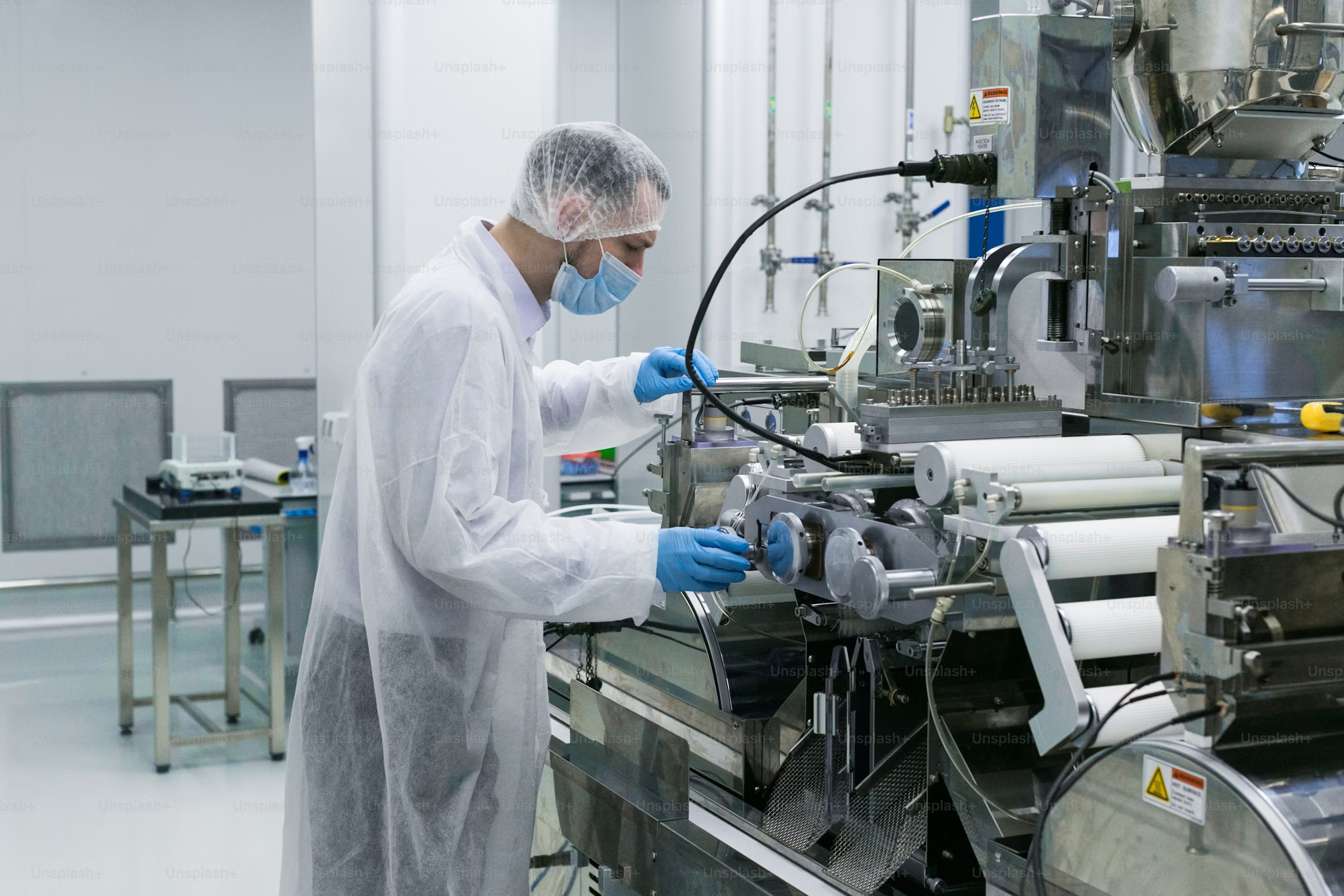
A new method of using light to scan the human body, developed by researchers at the University of St Andrews, could result in less intrusive and more effective diagnosis for patients. The work is the result of a collaboration between researchers from the Schools of Physics and Astronomy, Biology, Medicine and the Scottish Oceans Institute at the University.
The new technique allows the light to be shaped so it can reach greater depths within biological tissue enabling high quality three-dimensional (3D) images to be acquired. It can also allow detailed 3D images of biological specimens to be made without dissection or having to rotate specimens and take multiple images which are then fused together.
Published in the journal Science Advances, the research shows that the new method adds value to two existing imaging techniques – Bessel beam based light-sheet microscopy and Airy beam based light-sheet microscopy.
Dr Jonathan Nylk of the School of Physics and Astronomy said: ‘We’ve recently discovered particular beam shapes that retain their shape when travelling through biological tissue. These beams, called Airy beams and Bessel beams resist the effects of scattering but they still become dimmer as they travel deeper, so it remains challenging to collect enough signal back through the tissue to form an image.
‘Now we show that these beams can be further enhanced to give us more control over their shape, such that they actually get brighter as they travel (propagate). When the increase in brightness (intensity) is matched with the decrease in brightness (intensity) when travelling through tissue, a strong signal and a clear image can still be acquired from deep within the sample.’
This latest research builds on previous advances in ‘light-sheet imaging’, in which a thin sheet of light cuts across the sample like a razor blade to section the sample ‘ but without actually cutting or damaging it. The use of curved Airy light-sheets was shown to give sharp images over a volume ten times larger than previously possible.
The new techniques are expected be useful, not only for light-sheet microscopy, but also for pushing the limits of a whole range of other optical imaging techniques.
It is hoped that the development will lead to improved understanding of biological development, cancer, and diseases such as Alzheimer, Parkinson, and Huntington’s that affect the human brain.
The paper ‘Light-sheet microscopy with attenuation-compensated propagation-invariant beams’ by Dr Jonathan Nylk, Dr Kaley McCluskey, Dr Miguel A. Preciado, Dr Michael Mazilu, Dr Zhengyi Yang, Prof Frank J. Gunn-Moore, Ms Sanya Aggarwal, Dr Javier A. Tello, Prof David E. K. Ferrier, and Prof Kishan Dholakia is published in the (April issue of Science Advances), available via http://advances.sciencemag.org/content/4/4/eaar4817.